Biology
-
Charts
Angiosperms
These hand-drawn charts on rexine offer a unique and artistic approach to visual learning. Measuring 68×100 cm, they provide ample space for detailed illustrations, making complex concepts easy to understand. Durable and flexible, these charts are perfect for classrooms, presentations, or as decorative educational tools.
BG13122 -
Charts
Seeds & Fruits
These hand-drawn charts on rexine offer a unique and artistic approach to visual learning. Measuring 68×100 cm, they provide ample space for detailed illustrations, making complex concepts easy to understand. Durable and flexible, these charts are perfect for classrooms, presentations, or as decorative educational tools.
BG13124 -
Charts
Roots, Stems, Leaves
These hand-drawn charts on rexine offer a unique and artistic approach to visual learning. Measuring 68×100 cm, they provide ample space for detailed illustrations, making complex concepts easy to understand. Durable and flexible, these charts are perfect for classrooms, presentations, or as decorative educational tools.
BG13126 -
Charts
Flower & Embryology
These hand-drawn charts on rexine offer a unique and artistic approach to visual learning. Measuring 68×100 cm, they provide ample space for detailed illustrations, making complex concepts easy to understand. Durable and flexible, these charts are perfect for classrooms, presentations, or as decorative educational tools.
BG13128 -
Charts
Histology
These hand-drawn charts on rexine offer a unique and artistic approach to visual learning. Measuring 68×100 cm, they provide ample space for detailed illustrations, making complex concepts easy to understand. Durable and flexible, these charts are perfect for classrooms, presentations, or as decorative educational tools.
BG13130 -
Charts
Human Anatomy Charts
These hand-drawn charts on rexine offer a unique and artistic approach to visual learning. Measuring 68×100 cm, they provide ample space for detailed illustrations, making complex concepts easy to understand. Durable and flexible, these charts are perfect for classrooms, presentations, or as decorative educational tools.
BG13145 -
Charts
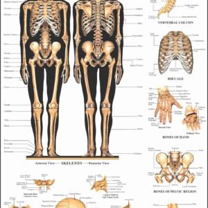
Human Anatomy
These vacuum-formed biological charts are made from heavy-duty plastic, offering deep relief for clear, tactile representation. Weatherproof and washable, they are built to withstand years of classroom use. With realistic colors and an aesthetically pleasing design, these charts provide valuable assistance in helping students understand biological concepts. They are also specially designed to be helpful for blind students, making learning more accessible.
Set of 11
(1) Skeleton
(2) Circulatory system
(3) Heart
(4) Nervous system
(5) Section of Brain
(6) Muscle
(7) Digestive system
(8) Respiratory system
(9) Ear
(10) Eye
(11) Kidneys.While size 25 x 35 cm is available in sets only, other sizes are available singly as well.
BG13147 -
Charts
Human Reproductive Organs
These vacuum-formed biological charts are made from heavy-duty plastic, offering deep relief for clear, tactile representation. Weatherproof and washable, they are built to withstand years of classroom use. With realistic colors and an aesthetically pleasing design, these charts provide valuable assistance in helping students understand biological concepts. They are also specially designed to be helpful for blind students, making learning more accessible.
Set of 8
(1) Male genital organs (front view)
(2) Male genital organs (sagittal view)
(3) How semen is formed
(4) First week of egg development and implantation
(5) Fertilization (cross section diagram of an ovary)
(6) Eight months old fetus
(7) Various stages of embryonic development
(8) Female genital organs (sagittal view)While size 25 x 35 cm is available in sets only, other sizes are available singly as well.
BG13149 -
Charts
Botany Series I
These vacuum-formed biological charts are made from heavy-duty plastic, offering deep relief for clear, tactile representation. Weatherproof and washable, they are built to withstand years of
classroom use. With realistic colors and an aesthetically pleasing design, these charts provide valuable assistance in helping students understand biological concepts. They are also specially designed to be helpful for blind students, making learning more accessible.Set of 8
(1) Root, stem and bud (A)
(2) Root, stem and bud (B)
(3) Leaves and their transformation
(4) Photosynthesis
(5) Flower and inflorescence (A)
(6) Flower and inflorescence (B)
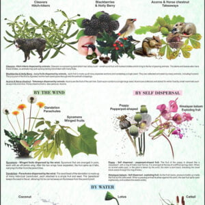
(7) Fruit and seeds (A)
(8) Fruit and seeds (B)While size 25 x 35 cm is available in sets only, other sizes are available singly as well.
BG13151 -
Charts
Botany Series II
These vacuum-formed biological charts are made from heavy-duty plastic, offering deep relief for clear, tactile representation. Weatherproof and washable, they are built to withstand years of
classroom use. With realistic colors and an aesthetically pleasing design, these charts provide valuable assistance in helping students understand biological concepts. They are also specially designed to be helpful for blind students, making learning more accessible.Set of 8
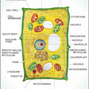
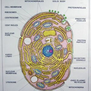
(1) Typical plant cell
(2) Plant Mitosis
(3) Meiosis
(4) DNA
(5) RNA
(6) Bacteria
(7) Spirogyra
(8) FunariaWhile size 25 x 35 cm is available in sets only, other sizes are available singly as well.
BG13153 -
Charts
Botany Series III
These vacuum-formed biological charts are made from heavy-duty plastic, offering deep relief for clear, tactile representation. Weatherproof and washable, they are built to withstand years of
classroom use. With realistic colors and an aesthetically pleasing design, these charts provide valuable assistance in helping students understand biological concepts. They are also specially designed to be helpful for blind students, making learning more accessible.Set of 8
(1) Fertilization
(2) T.S. Dicot leaf
(3) T.S. Monocot leaf
(4) T.S. Dicot root
(5) T.S. Monocot root
(6) T.S. Dicot stem
(7) T.S. Monocot stem
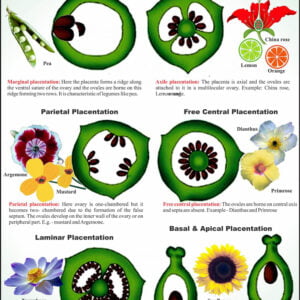
(8) Types of placentationWhile size 25 x 35 cm is available in sets only, other sizes are available singly as well.
BG13155 -
Charts
Botany Series IV
These vacuum-formed biological charts are made from heavy-duty plastic, offering deep relief for clear, tactile representation. Weatherproof and washable, they are built to withstand years of
classroom use. With realistic colors and an aesthetically pleasing design, these charts provide valuable assistance in helping students understand biological concepts. They are also specially designed to be helpful for blind students, making learning more accessible.Set of 8
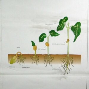
(1) Plant kingdom
(2) Germination of sunflower and maize
(3) Germination of bean
(4) Yeast and pea
(5) Rhizopus and Mucor
(6) Fern
(7) Insectivorous plants
(8) MalvaceaeWhile size 25 x 35 cm is available in sets only, other sizes are available singly as well.
BG13157 -
Charts
Zoology Series I
These vacuum-formed biological charts are made from heavy-duty plastic, offering deep relief for clear, tactile representation. Weatherproof and washable, they are built to withstand years of
classroom use. With realistic colors and an aesthetically pleasing design, these charts provide valuable assistance in helping students understand biological concepts. They are also specially designed to be helpful for blind students, making learning more accessible.Set of 8
(1) Typical animal cell
(2) Animal mitosis
(3) Hydra
(4) Paramecium
(5) Amoeba proteus
(6) Hookworm
(7) Tapeworm
(8) EarthwormWhile size 25 x 35 cm is available in sets only, other sizes are available singly as well.
BG13159 -
Charts
Zoology Series II
These vacuum-formed biological charts are made from heavy-duty plastic, offering deep relief for clear, tactile representation. Weatherproof and washable, they are built to withstand years of
classroom use. With realistic colors and an aesthetically pleasing design, these charts provide valuable assistance in helping students understand biological concepts. They are also specially designed to be helpful for blind students, making learning more accessible.Set of 8
(1) Euglena
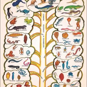
(2) Animal kingdom
(3) L.H. of frog(development)
(4) L.H. of frog(metamorphosis)
(5) L.H. of mosquito
(6) Epithelial & connective tissues
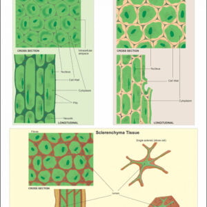
(7) Simple and complex tissues
(8) Rabbit dissection and skeletonWhile size 25 x 35 cm is available in sets only, other sizes are available singly as well.
BG13161 -
Charts
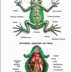
Zoology Series III
These vacuum-formed biological charts are made from heavy-duty plastic, offering deep relief for clear, tactile representation. Weatherproof and washable, they are built to withstand years of
classroom use. With realistic colors and an aesthetically pleasing design, these charts provide valuable assistance in helping students understand biological concepts. They are also specially designed to be helpful for blind students, making learning more accessible.Frog
Set of 8
(1) Frog anatomy and skeleton
(2) Frog digestive system
(3) Frog circulatory system
(4) Frog respiratory system
(5) Frog nervous system
(6) Frog reproductive system, male
(7) Frog reproductive system, female
(8) Frog heartWhile size 25 x 35 cm is available in sets only, other sizes are available singly as well
BG13163 -
Charts
Zoology Series IV
These vacuum-formed biological charts are made from heavy-duty plastic, offering deep relief for clear, tactile representation. Weatherproof and washable, they are built to withstand years of
classroom use. With realistic colors and an aesthetically pleasing design, these charts provide valuable assistance in helping students understand biological concepts. They are also specially designed to be helpful for blind students, making learning more accessible.Rat
Set of 8
(1) Rat anatomy,dissection showing internal organs (female)
(2) Rat digestive system
(3) Rat circulatory system
(4) Rat respiratory system
(5) Rat excretory system
(6) Rat reproductive system (male)
(7) Rat reproductive system (female)
(8) Rat brain & heartWhile size 25 x 35 cm is available in sets only, other sizes are available singly as well
BG13165 -
Charts
Zoology Series V
These vacuum-formed biological charts are made from heavy-duty plastic, offering deep relief for clear, tactile representation. Weatherproof and washable, they are built to withstand years of
classroom use. With realistic colors and an aesthetically pleasing design, these charts provide valuable assistance in helping students understand biological concepts. They are also specially designed to be helpful for blind students, making learning more accessible.Set of 8
(1) Life History of Honey Bee
(2) Life History of Silkworm
(3) Life History of House Fly
(4) Malarial Parasite (Plasmodium)
(5) Cockroach, circulatory & nervous systems
(6) Cockroach, external features
(7) Cockroach, digestive & respiratory systems
(8) Earthworm, circulatory & excretory systemsWhile size 25 x 35 cm is available in sets only, other sizes are available singly as well.
BG13167 -
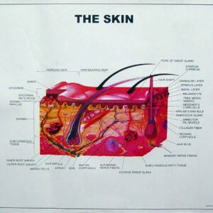
Charts
Human Skin
These vacuum-formed biological charts are made from heavy-duty plastic, offering deep relief for clear, tactile representation. Weatherproof and washable, they are built to withstand years of classroom use. With realistic colors and an aesthetically pleasing design, these charts provide valuable assistance in helping students understand biological concepts. They are also specially designed to be helpful for blind students, making learning more accessible.
BG13169